quinta-feira, 19 de setembro de 2013


CURSO DE ATUALIZAÇÃO EM BANDAGEM ELÁSTICA TERAPÊUTICA


Ministrante:

- Dr. Carlos Alberto Vieira de Melo - 13953-F

- Fisioterapeuta

- Pós - graduado em Fisioterapia Desportiva

- Mestrando – Mestrado da Saúde, Educação e Ambiente – UNIPLI

- Graduando – Gestão Hospitalar – UNESA

- Prof. Do curso de Fisioterapia Unesa – Fisioterapia Desportiva, Aparelho Locomotor, Avaliação Cinético Funcional.

- Prof. Do curso de Enfermagem da Unesa-  Filosofia 

- Prof. Da Pós-graduação em Enfermagem da Unesa- Ética na Emergência

- Consultor Esportivo – International School Sport Nutrition and Human      Performance,

- Formado no método Kabath, Mulligan, Spryral taping, Bandagem funcional, Kinesio taping, Terapia Manual (método australiano), Bola Suíça, Theraband, Sistema Raith, Crochetagem.

- Formado no método Wii e X Box 360 Jogos para Reabilitação- Hospital das Clínicas – SP.

- Formado na CIF Brasil.

- Formado no curso livre de Filosofia da Educação – ESAB

- Formado no curso livre de Filosofia – FGV

- Formado no curso de Direito na Saúde – Portal Educação.


Data: 09 de Novembro DE 2013 na Clínica CEFI


O CURSO É DESTINADO AS PESSOAS QUE JÁ FIZERAM O CURSO.


NESTE ENCONTRO VAMOS ABORDAR NOVAS TÉCNICAS E DISCUTIR OS RESULTADOS ENCONTRADO NA PRÁTICA.


Valor do curso:


100,00 Reais.


TEL: 0XX 22 99625676.


domingo, 28 de outubro de 2012


Gostaria de agradecer a todos os envolvidos no Congresso de Fisioterapia da Universidade Estácio de Sá  à Homenagem a mim conferida. Não tenho palavras para agradecer tamanha homenagem, mais ainda, ter uma sala com o meu nome é  muito gratificante. Isso  me obriga a ter ainda mais responsabilidade, não só com a minha profissão, mais principalmente com  meus clientes e alunos.
O meu muito obrigado.

sábado, 25 de fevereiro de 2012

Pesquisa científica ou genocídio programado?


Prezados amigos (as), estou reproduzindo na integra uma parte do livro Cobaias Humanas do autor Andrew Goliszek no que se refere ao futuro com pesquisas na área da medicina molecular.
É sabido que as pesquisas são uma parte importante para o crescimento, desenvolvimento e melhoria da qualidade de vida de uma nação, o que me preocupa qual  é o preço que nós estamos pagando em pró do desenvolvimento desenfreado das nações mundo afora.  

Segue um pequeno trecho do que pode acontecer em termos de pesquisa em Medicina Molecular até o ano 2020.

Antes da metade deste século, nada que os escritores de ficção cientifica pudessem ter sonhado irá se comparar àquilo que a ciência e a medicina terão de fato realizado. Para se ter idéia, no Laboratório Nacional de Los Alamos do governo dos EUA, no novo México, o uso de supercomputadores e de análise avançada de dados está acelerando o desenvolvimento de vacinas e tratamentos medicamentosos que resistem a mudanças evolutivas nos patógenos. Juntamente com um consórcio de instituições acadêmicas e empresas comerciais farmacêuticas e biotecnológicas, os pesquisadores de Los Alamos desenvolvem modelos computacionais que estimularão as seqüências moleculares para moldar drogas projetadas para combater doenças.
A verdadeira guerra biológica deste século ocorrerá nos campos de batalha hospitalares e nos consultórios dos médicos, onde cepas letais de micróbios se tornarão tão inócuas quanto um leve resfriado.
Algumas das ferramentas de modelagem por computador foram inicialmente desenvolvidas para analisar vírus que sofriam  mutações rápidas, como o HIV, e então, mais tarde, expandiram-se para incluir o novo campo da “diversidade molecular”. Uma área altamente promissora da genética, a finalidade da diversidade molecular é controlar e direcionar a evolução através da criação de um ambiente em que as moléculas evoluem através de seleção artificial, em vez de seleção natural aleatória.  De acordo com o Dateline Los Alamos, com a geração de bilhões de moléculas diversas de DNA, RNA, proteínas e outras moléculas orgânicas aleatoriamente para ver qual se sai melhor em se ajustar a um receptor em, digamos, um envoltório viral, os melhores candidatos serão identificados e reproduzidos com mutações para acelerar uma versão laboratorial da evolução. Esse processo de seleção artificial permite que os cientistas desenvolvam e testem um enorme número de variantes em questão de horas e gerem um tremendo número de dados de diversidade molecular.
O Laboratório Los Alamos, é o laboratório que estuda a estrutura molecular de cepas do HIV, e este laboratório foi escolhido pela OMS para atuar nos países de Uganda, Ruanda, Tailândia e Brasil. Este laboratório também tem um projeto para gerar um banco de dados do vírus do papiloma humano, a principal doença sexualmente transmitida do mundo.
Com certeza com o maior conhecimento sobre nossos genes certamente salvará vidas, isso invariavelmente levará a uma sociedade na qual escolheremos quem vive e quem morre, com base no DNA. Hoje já existe visita a laboratório médico para examinar o próprio DNA para verificar a presença de uma doença ou problema que possam a aparecer.
O lado negro disso tudo é que os indivíduos que, do contrario poderiam nascer e se transformar em grandes pensadores, lideres mundiais, cientistas e empreendedores poderão acabar sendo eliminados porque um teste genético pré – natal revelou uma doença. Mesmo os testes genéticos “preditivos”, que atribuem uma probabilidade de contrair uma doença com base na historia familiar, seriam usados para pôr fim a uma gravidez que poderia ou não resultar na doença.
Avaliar os riscos e, então derrotar as probabilidades de passar genes defeituosos aos descendentes torna-se-à  então o objetivo do jogo. A eugenia, que horrorizou tantos no passado, realmente não está tão longe assim de se tornar a prática convencional para muitos casais em busca de seu bebê perfeito.

um grande abraço.

quarta-feira, 11 de janeiro de 2012

Exposições em Museu

Prezados amigos(as), dando continuidade ao que está acontecendo em termos de cultura no mundo, estou colocando alguns avisos sobre visitas em museus.
 No Brasil acontecerá no final do mês Um dos mais importantes eventos do calendário oficial do Momento Itália - Brasil, a exposição “Modigliani: Imagens de uma vida” será inaugurada em 31 de janeiro no MNBA, com  obras nunca antes expostas na America Latina. Para o público a mostra abre dia 1º de fevereiro.
Serão exibidas 12 pinturas e 5 esculturas originais, além de obras e documentos, fotos,  desenhos, diários e manuscritos de Modigliani e de importantes artistas da sua época, num total de 230 peças. Com isso o público vai ter acesso a um rico panorama da vida artística parisiense e italiana do século XX, distribuídas em cinco salas do MNBA

aproveitem as férias e divirtam-se.


No The Metropolitan Museum of Art, New York, está acontecendo a  exposição  "retrato renascentista de Donatello para Bellini "

21 de dezembro de 2011 - 18 de março, 2012

 
Tem sido dito que a Renascença testemunhou a redescoberta do indivíduo. De acordo com esta noção, no início da Renascença a Itália também sediou a primeira era grande do retrato na Europa. Retrato assumiu uma nova importância, se era para gravar as características de um membro da família para as gerações futuras, celebrar um príncipe ou um guerreiro, exaltar a beleza de uma mulher, ou tornar possível a troca de uma semelhança entre amigos. Esta exposição reunirá cerca de 160 obras, de artistas como Donatello, Filippo Lippi, Botticelli, Verrocchio, Ghirlandaio, Pisanello, Mantegna, Bellini Giovanni, e da Antonello de Messina, e nos meios de comunicação que vão desde iluminação e pintura manuscrito a escultura em mármore e bronze , atestando a nova moda e usa do retrato no século XV na Itália.
Durante o Renascimento, os artistas que trabalham em Florença, Veneza, e os tribunais da Itália criou retratos magníficos das pessoas ao seu redor, desde chefes de Estado e igreja para patronos, acadêmicos, poetas e artistas, concentrando-se pela primeira vez na produção reconhecíveis semelhanças e as expressões da personalidade. O rápido desenvolvimento do retrato estava ligado intimamente ao Renascimento a sociedade ea política, os ideais do indivíduo, e conceitos de beleza. O objeto pode ter sido para comemorar um evento-a significativa casamento, a morte, a adesão a uma posição de poder ou pode ter sido para gravar as características de um membro estimado da família para as gerações futuras.
Apresentando muitos raros empréstimos internacionais, esta exposição irá apresentar um levantamento inédito do período e fornecer novas pesquisas e insights sobre o início da história do retrato. Será dividido em três seções e terá uma duração de um período de oito décadas. Início em Florença, onde os retratos independente apareceu pela primeira vez em abundância, ela move-se para os tribunais de Ferrara, Mântua, Bolonha, Milão, Urbino, Nápoles e Roma papal, e termina em Veneza, onde a tradição do retrato afirmou-se surpreendentemente no final do século .

Quem tem dinheiro e disponibilidade é uma boa pedida.


Vesalius, Andreas

Prezados amigos, como o meu blog é bem diversificado, estou abrindo um espaço para divulgar as grandes obras de artes de vários artistas de varias épocas e séculos.
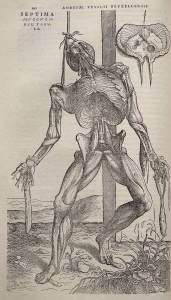
Estou começando com um grande artista  Sr. Andreas Vesalius, que contribuiu muito para o ensino na área da Anatomia.
Ao lado estamos vendo uma de suas obras publicada em 1543 " Dissecação do corpo humano em pose alegórica".
Obra Renascentista.


Andreas Vesalius (1514-1564) nasceu pouco antes da meia-noite do dia 31 de dezembro de 1514. Tanto seu bisavô quanto o avô haviam sido médicos, e o pai era o boticário do imperador Maximiliano I, da família Habsburg. Incentivado pelo pai a seguir a profissão da família, Vesalius cursou primeiro a Universidade de Louvain, perto de Bruxelas, na Bélgica, e depois começou a estudar medicina na Universidade de Paris.
Nas aulas, os professores liam livros da Antigüidade sobre personagens como Galeno, o anatomista grego, enquanto seus assistentes conduziam dissecações desastradas. Vesalius dizia que conseguiria aprender mais com um açougueiro no mercado de carnes. Em 1536, ele voltou a Louvain, onde aproveitou uma chance de estudar o esqueleto humano. Encontrou o cadáver de um ladrão pendurado em uma forca nas imediações da cidade. Pássaros haviam comido toda a carne, limpando os ossos do esqueleto. Vesalius o desmembrou e estudou até conhecer todos os ossos, até mesmo com os olhos vendados.
Em 5 de dezembro de 1537, Vesalius se formou em medicina pela Universidade de Pádua, perto de Veneza e tornou-se professor de anatomia no dia seguinte. Segundo a promessa que havia feito a si mesmo, ensinava anatomia conduzindo ele próprio as dissecações. As aulas de Vesalius tornaram-se imensamente populares. Os alunos se amontoavam na sala para observar suas habilidosas dissecações. De forma a mostrar pequenos detalhes a grandes platéias e para ter alguma coisa que lhe ajudasse nas palestras quando não havia cadáveres disponíveis, Vesalius desenhou mapas dos órgãos humanos, que pendurou na sala de aula. Alguns desses mapas foram plagiados e publicados sem sua permissão. Ele publicou o restante dos mapas com um manual de anatomia para completá-los.
O sucesso do manual o incentivou a publicar De Humani Corpus Fabrica (A Matéria do Corpo Humano). Esta grande obra foi publicada em 1543 em sete volumes, com descrições detalhadas e 300 ilustrações. Vesalius escolheu Jan Stephen van Calcar, um jovem aluno do artista Ticiano, para aperfeiçoar seus esboços de forma que os pontos essenciais pudessem ser compreendidos rapidamente. Os desenhos mostravam o corpo humano em poses naturais. No livro, Vesalius detalhou suas descobertas e corrigiu mais de 200 erros que havia encontrado nos livros de Galeno. Ele também revelou que Galeno havia baseado suas teorias de anatomia em dissecações de animais.
O livro de Vesalius gerou forte oposição. O anatomista principal de Paris, Jacques Sylvius, o atacou, dizendo se um insulto a Galeno. Vesalius tinha 28 anos de idade quando publicou A Matéria do Corpo Humano. Depois de defender o livro por um ano, ele deixou o magistério e as pesquisas de anatomia. Daquele ponto em diante, trabalhou como médico na corte dos Habsburg, primeiro para o imperador Carlos V, filho de Maximiliano, e depois para o filho de Carlos, Felipe II, em Madri.
Com 50 anos de idade, Vesalius viajou a Jerusalém, não se sabe ao certo por quê. A peregrinação pode ter sido feita para calar os críticos que o acusavam de falta de piedade. Tragicamente, na viagem de volta, Vesalius morreu em um naufrágio perto da ilha grega de Zakinthos.
Fonte de pesquisa:
www.historia.com.br


Magnetic Resonance Imaging: Generating a New Pulse in the Physical Therapy Profession

Prezados colegas, a cada dia estamos sendo testados nas nossas profissões. Com as novas tecnologias de ponta, muitos dos problemas  patológicos e principalmente das dores sem causas pré- estabelecidas  que exigiam de nós um conhecimento quase que transcendental, dificultando aos profissionais de saúde em fechar um diagnóstico preciso, sendo na sua grande maioria fechado apenas com base na anamnese e no exame clínico.  Hoje com a qualidade dos aparelhos de imagens, os profissionais de saúde têm encontrado mais facilidade em confirmar o que tem encontrado na anamnese e nos exames clínicos e confirmando com os exames de imagens. A Ressonância Magnética é um bom exemplo desse tipo de exame, que hoje facilita bastante aos profissionais a fecharem um diagnóstico mais coerente, principalmente os de causas pré-estabelecidas.
Por outro lado estamos vendo cada vez mais os profissionais abandonarem a prática da avaliação seja ela clínica ou funcional; não podemos nos esquecer que os exames de imagens por natureza apresentam uma característica auxiliadora, o que isso quer dizer, é que o exame de imagem é secundário ao exame clínico ou funcional, lógico dependendo de cada caso, pode ser que eles tenham em determinados momentos um caráter primário.
O fisioterapeuta dentro das suas funções tem a total liberdade de solicitar exames que venham a elucidar  e ajudar a fechar o seu fisiodiagnóstico, porém  a dificuldade desse profissional em lidar com exames de imagem ainda é muito grande, exigindo de nós um compromisso maior em aprender a entender um exame desse porte. Acredito que neste momento e com a exigência do mercado, que tem exigido cada vez mais profissionais qualificados, as Universidades que oferecem o curso de Fisioterapia deveriam se preocupar mais com esta parte da matéria. Como é de costume, lendo um artigo na revista JOSPT encontrei um artigo que fala sobre o avanço da fisioterapia neste campo do conhecimento.
Espero que seja proveitoso para vocês.

 
Magnetic Resonance Imaging: Generating a New Pulse in the Physical Therapy ProfessionJAMES M. ELLIOTT, PT, PhD1J Orthop Sports Phys Ther 2011;41(11):803-805. doi:10.2519/jospt.2011.0109
I
n 2003, the Board of Directors of the American Physical Therapy Association (APTA) published a position statement (BOD-P03-03-12-28) promoting the goal that all physical therapists hold the privileges of autonomous practice. In 2006, the House of Delegates of the APTA moved forward on the same topic with their position statement (HOD P06-06-18-12), reaffirming the concept from the
BOD that "Autonomous physical therapist practice is characterized by independent, self-determined professional judgment and action." The statement further defined the 4 expected privileges of the autonomous physical therapist practitioner by the year 2020:
1. Direct and unrestricted access: the physical therapist has the professional capability and ability to provide to all individuals the physical therapy services they choose without legal, regulatory, or payer restrictions.
2. Professional ability to refer to other healthcare providers: the physical therapist has the professional capability and ability to refer to others in the healthcare system for identified or possible medical needs beyond the scope of physical therapist practice.
3. Professional ability to refer to other professionals: the physical therapist has the professional capability and ability to refer to other professionals for identified patient/client needs beyond the scope of physical therapist practice.
4. Professional ability to refer for diagnostic tests: the physical therapist has the professional capability and ability to refer for diagnostic tests that would clarify the patient/client situation and enhance the provision of physical therapy services.
Professional Ability to Refer for Diagnostic Tests
While each of the 4 privileges offer an exciting, and mostly measurable, glimpse into autonomous practice, the ability to refer for diagnostic tests is particularly relevant to the topic of this special issue on magnetic resonance imaging (MRI). As a doctoring profession, one could argue that the ability to refer a patient for MRI (and other imaging applications) must be available to the direct-access physical therapist. Though the prescription for and use of MRI has not typically been considered within the scope of physical therapist practice, as others have detailed,
 READINESS: WHAT DOES THE EVIDENCE SAY?
P
hysical therapists undergo extensive didactic and clinical preparation; they possess excellent diagnostic skills, and are well versed in current best diagnostic and treatment-based evidence.4 Physical therapists are well suited to become key members of an interdisciplinary healthcare team that makes early decisions towards retarding or preventing the development of long-term physical impairments second
1
Assistant Professor, Department of Physical Therapy and Human Movement Sciences, Feinberg School of Medicine, Northwestern University, Chicago, IL; Honorary Senior Fellow, School of Health and Rehabilitation Sciences, The University of Queensland, Brisbane, Australia
journal of orthopaedic
& sports physical therapy | volume 41 | number 11 | november 2011 | 803
ary to injury and/or disease processes. A system that places physical therapists as first-line providers for patients with painful neuromusculoskeletal conditions may help to realize such an outcome.22 and The University of Wisconsin Hospital and Clinics.2 It must be emphasized that the physical therapists in these settings have undergone extensive advanced training in evaluating, diagnosing, and treating patients’ conditions in a direct-access setting, and in ordering imaging to aid in diagnosis when necessary. These examples provide evidence that an advanced training program can effectively establish the primary care scope of the physical therapist. But just as importantly, we must add that the data emerging from these model centers may not yet be generalizable to our national or worldwide physical therapist communities.19 In an attempt to reduce specialty care costs (eg, ordering of imaging) for patients with low back pain, changes in the clinical decision pathway were proposed and included implementation of an evidenced-based protocol with physical therapists up front. After 1 year, the model yielded favorable results. The number of ordered MRI exams and total cost per episode of care were both reduced. Furthermore, the initiation of direct-access physical therapy resulted in fewer patients transiting to the chronic pain center. While further research is required before definitive conclusions can be drawn, this example provides strong foundation for welcoming physical therapists who are well versed in the implementation of appropriate imaging guidelines, as front-line providers.
Health systems in which physical therapists are specifically empowered to function as first-line providers include the US Military, Kaiser Permanente Northern California, the Department of Veterans Affairs Salt Lake City Health Care Systems,
Another example of physical therapists functioning under expanded primary care privileges is the 2004 pilot project from the Virginia Mason Medical Center in Seattle, WA.
 TOWARDS A NEW NORMATIVE MODEL FOR PHYSICAL THERAPIST PRACTICE
I
An important personal context for this special issue is my belief that such interdisciplinary professional collaboration, at least with respect to MRI, can be further cultivated by physical therapists who seek formal or informal opportunity to gain an improved working knowledge of (1) the complexities of MRI,
ncorporation of didactic content on diagnostic imaging (eg, radiography, computed tomography, ultrasound, MRI) has become the educational norm for entry-level and transition Doctor of Physical Therapy degree students. Additionally, there are a number of professional development opportunities offered through advanced clinical practice courses by the APTA. Clinicians and researchers in our field have published and continue to publish seminal and oftentimes multidisciplinary works related to imaging.6,8,9,12-16,23,24,26-28 Our journals continue to publish more imaging-related studies/cases, and our professional conferences encourage and accept poster and platform submissions that feature imaging. Finally, the recent development of the Special Interest Group on Imaging within the Orthopedic Section of the APTA is another important contribution to developing practice competencies, reducing practice variability, fostering imaging curriculum in physical therapist education, and promoting the role of the physical therapist, as a primary care team member, in judicious referral for imaging procedures. These all represent a great start for informing the entire physical therapist community and other stakeholders (medical practitioners, administrators, insurers, legislators, and the public) regarding the evolving important role of imaging within the scope of physical therapy practice. However, to realize the maximum potential and success of such a model, collegial collaboration with others inside and outside the physical therapy profession is imperative. This collaborative approach is consistent with the definition of professional autonomy, not deviant from it.21 (2) the issues related to patient and operator safety when working in an MRI environment,10 (3) determining the best sequences for identifying pathology,11,29,30 (4) emerging evidence that highlights advanced imaging applications,1,5 (5) continued vigilance regarding overutilization of MRI services,17 and, finally, (6) a history of physical therapy and imaging.4
This special issue contains a collection of manuscripts, authored by multidisciplinary teams, that address each of the aforementioned topics. As such, it is highly relevant for all clinicians involved in managing patients with neuromusculoskeletal disorders, because MRI may play a significant role in helping to make more informed clinical decisions. More importantly, we hope that the information presented here will inspire readers to seek and embrace opportunities for fostering and maintaining collegial collaborations inside and outside the professional boundaries of physical therapy.

POUNDING HOME THE TRUTH
I
Finally, I would like to thank all the authors for their contribution to this issue and also the multidisciplinary team of reviewers for their expertise, time, and effort in providing peer review.
 
804
| november 2011 | volume 41 | number 11 | journal of orthopaedic & sports physical therapy
 
REFERENCES
1. Beattie PF. Diffusion-Weighted Magnetic Resonance Imaging of the Musculoskeletal System: An Emerging Technology With Potential to Impact Clinical Decision Making. J Orthop Sports Phys Ther. 2011;41:887-895. http://dx.doi.org/10.2519/jospt.2011.3744 2. Boissonnault WG, Badke MB, Powers JM. Pursuit and implementation of hospital-based outpatient direct access to physical therapy services: an administrative case report. Phys Ther. 2010;90:100-109. http://dx.doi.org/10.2522/ptj.20080244 3. Boissonnault WG, Goodman C. Physical therapists as diagnosticians: drawing the line on diagnosing pathology. J Orthop Sports Phys Ther. 2006;36:351-353. http://dx.doi.org/10.2519/jospt.2006.0107 4. Boyles R, Gorman I, Pinto D, Ross M. Future physical therapist practice and the role of diagnostic imaging. J Orthop Sports Phys Ther. 2011;41:829-837. http://dx.doi.org/10.2519/jospt.2011.3556 5. Cagnie B, Elliott J, O’Leary S, D’hooge R, Dickx N, Danneels L. Muscle functional MRI as an imaging tool to evaluate muscle activity. J Orthop Sports Phys Ther. 2011;41:896-903. http://dx.doi.org/10.2519/jospt.2011.3586 6. Cagnie B, O’Leary S, Elliott J, Peeters I, Parlevliet T, Danneels L. Pain-induced changes in the activity of the cervical extensor muscles evaluated by muscle functional magnetic resonance imaging. Clin J Pain. 2010;27:392-397. http://dx.doi.org/10.1097/AJP.0b013e31820e11a2 7. Davenport TE, Kulig K, Resnik C. Diagnosing pathology to decide the appropriateness of physical therapy: what’s our role? J Orthop Sports Phys Ther. 2006;36:1-2. http://dx.doi.org/10.2519/jospt.2006.0101 8. Deyle GD. Musculoskeletal imaging in physical therapist practice. J Orthop Sports Phys Ther. 2005;35:708-721. http://dx.doi.org/10.2519/jospt.2005.2034 9. Dickx N, D’Hooge R, Cagnie B, Deschepper E, Verstraete K, Danneels L. Magnetic resonance imaging and electromyography to measure lumbar back muscle activity. Spine (Phila Pa 1976). 35:E836-842. http://dx.doi.org/10.1097/BRS.0b013e3181d79f02 10. Durbridge G. Magnetic resonance imaging: fundamental safety issues. J Orthop Sports Phys Ther. 2011;41:820-828. http://dx.doi.org/10.2519/jospt.2011.3906 11. Elliott JM, Flynn T, Press J, Al-Najjar A, Nguyen B, Noteboom J. The pearls and pitfalls of magnetic resonance imaging for the spine. J Orthop Sports Phys Ther. 2011;41:848-860. http://dx.doi.org/10.2519/jospt.2011.3636 12. Elliott JM, Jull G, Noteboom JT, Darnell R, Galloway G, Gibbon WW. Fatty infiltration in the cervical extensor muscles in persistent whiplash-associated disorders: a magnetic resonance imaging analysis. Spine (Phila Pa 1976). 2006;31:E847-855. http://dx.doi.org/10.1097/01.brs.0000240841.07050.34 13. Elliott JM, Pedler A, Beattie P, McMahon K. Diffusion-weighted magnetic resonance imaging for the healthy cervical multifidus: a potential method for studying neck muscle physiology following spinal trauma. J Orthop Sports Phys Ther. 2010;40:722-728. http://dx.doi.org/10.2519/jospt.2010.3423 14. Elliott JM, Pedler A, Kenardy J, Galloway G, Jull G, Sterling M. The temporal development of fatty infiltrates in the neck muscles following whiplash injury: an association with pain and posttraumatic stress. PLoS One. 6:e21194. http://dx.doi.org/10.1371/journal.pone.0021194 15. Elliott JM, Sterling M, Noteboom JT, Darnell R, Galloway G, Jull G. Fatty infiltrate in the cervical extensor muscles is not a feature of chronic, insidious-onset neck pain. Clin Radiol. 2008;63:681-687. http://dx.doi.org/10.1016/j.crad.2007.11.011 16. Elliott JM, Pedler AR, Cowin G, Sterling M, McMahon K. Spinal cord metabolism and muscle water diffusion in whiplash. Spinal Cord. 2011 Mar 8. Epub ahead of print. http://dx.doi.org/10.1038/sc.2011.17 17. Flynn T, Smith B, Chou R. Appropriate use of diagnostic imaging in low back pain: a reminder that unnecessary imaging may do as much harm as good. J Orthop Sports Phys Ther. 2011;41:838-846. http://dx.doi.org/10.2519/jospt.2011.3618 18. Fritz J, Flynn TW. Autonomy in physical therapy: less is more. J Orthop Sports Phys Ther. 2005;35:696-698. http://dx.doi.org/10.2519/jospt.2005.0111 19. Fuhrmans V. A novel plan helps hospital wean itself off pricey tests. Available at: http://online.wsj.com. Accessed September 19, 2011, 2007. 20. Jarvik JG, Hollingworth W, Martin B, et al. Rapid magnetic resonance imaging vs radiographs for patients with low back pain: a randomized controlled trial. JAMA. 2003;289:2810-2818. http://dx.doi.org/10.1001/jama.289.21.2810 21. McMahon K, Cowin G, Galloway G. Magnetic resonance imaging: the underlying principles. J Orthop Sports Phys Ther. 2011;41:806-819. http://dx.doi.org/10.2519/jospt.2011.3576 22. Murphy BP, Greathouse D, Matsui I. Primary care physical therapy practice models. J Orthop Sports Phys Ther. 2005;35:699-707. http://dx.doi.org/10.2519/jospt.2005.2167 23. O’Leary S, Cagnie B, Reeve A, Jull G, Elliott JM. Is there altered activity of the extensor muscles in chronic mechanical neck pain? A functional magnetic resonance imaging study. Arch Phys Med Rehabil. 2011;92:929-934. http://dx.doi.org/10.1016/j.apmr.2010.12.021 24. Patten C, Meyer RA, Fleckenstein JL. T2 mapping of muscle. Semin Musculoskelet Radiol. 2003;7:297-305. http://dx.doi.org/10.1055/s-2004-815677 25. Ross MD, Boissonnault WG. Red flags: to screen or not to screen? J Orthop Sports Phys Ther. 2010;40:682-684. http://dx.doi.org/10.2519/jospt.2010.0109 26. Ross MD, Cheeks JM. Clinical decision making associated with an undetected odontoid fracture in an older individual referred to physical therapy for the treatment of neck pain. J Orthop Sports Phys Ther. 2008;38:418-424. http://dx.doi.org/10.2519/jospt.2008.2687 27. Ross MD, Cheeks JM. Undetected hangman’s fracture in a patient referred for physical therapy for the treatment of neck pain following trauma. Phys Ther. 2008;88:98-104. http://dx.doi.org/10.2522/ptj.20070033 28. Segal RL. Use of imaging to assess normal and adaptive muscle function. Phys Ther. 2007;87:704-718. http://dx.doi.org/10.2522/ptj.20060169 29. Strudwick M, Anderson S, Dimmick S, Saltzman M, Hsu W. Pearls and Pitfalls of Magnetic Resonance Imaging of the Upper Extremity. J Orthop Sports Phys Ther. 2011;41:861-872. http://dx.doi.org/10.2519/jospt.2011.3833 30.

journal of orthopaedic & sports physical therapy | volume 41 | number 11 | november 2011 | 805

Tall M, Thompson A, Greer B, Campbell S. The Pearls and Pitfalls of Magnetic Resonance Imaging of the Lower Extremity. J Orthop Sports Phys Ther. 2011;41:873-886. http://dx.doi.org/10.2519/jospt.2011.3713
n closing, as an old legal adage states: If you have the truth on your side, pound the truth. If you have the facts on your side, pound the facts. And if you have neither, pound the table. For today’s (and tomorrow’s) physical therapist, it is important to realize the responsibility that comes with the complex challenges of referral for imaging and other diagnostic privileges. As such, let’s work in an interdisciplinary environment towards discovering new facts that reveal new truths regarding physical therapists as front-line providers. Our field has and will continue to overcome professional challenges, but we must not be guilty of pounding the table. Let’s gather the facts and stick to the truth.t
 
3,7,8,18,22,28 this may be the perfect time to challenge the obstacles precluding the obtainment of such diagnostic privileges and to specifically provide perspective on why and how such a specialized privilege could positively impact the provision of physical therapy services and patient outcomes.8,28 Physical therapists can play a crucial primary-care role through participation in models based on diagnostic and patient management algorithms3,7,18,25 to (1) reduce practice variation,18 and (2) help to ensure that patients understand both the necessity for appropriate imaging and appreciate the potential negative consequences of unnecessary imaging.17,20 An expected obligation of such autonomy, however, is accountability. Is our entire profession adequately prepared to accept the role of, or the responsibilities associated with, referral for imaging privileges?